The Future of Regeneration and Cartilage Repair with Stem Cells
At Cellular Performance Institute (CPI), we pride ourselves on being at the forefront of cutting-edge medical technology. The latest milestone in our journey is launching a groundbreaking product: ChondRX™.
This revolutionary product contains chondrocytes, a cartilage-like cell fundamental to the health and repair of our joints. In combination with our advanced Hypoxic Mesenchymal Stem Cells (Hypo-MSCs) and a state-of-the-art 3D cellular matrix scaffold, ChondRX™ signifies a monumental leap in regenerative medicine.
Understanding Chondrocyte Cells for Cartilage Repair
Before we delve into the benefits and uniqueness of ChondRX™, it’s essential to understand what chondrocytes are and why they are so crucial to our health. Chondrocytes are specialized cells that form and maintain cartilage, a flexible and resilient tissue that supports various structures in our body, such as the ear, nose, and joints [1]. Among their many responsibilities, chondrocytes are known for producing key components of the cartilage matrix, including collagen, the primary structural protein in our bodies.
Collagen plays a central role in maintaining and repairing various tissues, including cartilage, skin, and bones. Chondrocytes generate a specific type of collagen, known as type II collagen, which provides strength and elasticity to our cartilage, allowing it to absorb shock and withstand physical stress [2]. It’s easy to see why these cells are so essential in addressing joint and disc regeneration!
The application of chondrocytes in regenerative medicine has been used previously. In a study by Brittberg et al., autologous chondrocytes (chondrocytes harvested from patients’ bodies) were successfully used to treat deep cartilage defects [3]. However, despite the success of autologous chondrocyte transplantation, this approach has limitations, such as potential harm to the donor site and limited cell volume [4]. In addition, patients using autologous Chondrocytes must also undergo surgery to remove cartilage and another more invasive surgery to implant the cells. Our allogeneic product does not need surgery, drastically reducing recovery time.
Hypoxic Mesenchymal Stem Cells (Hypo-MSCs): Potency Amplified
At CPI, we harness the power of Hypo-MSCs, a more advanced and potent mesenchymal stem cell (MSC) type. Unlike normoxic MSCs that are cultured under normal oxygen conditions, Hypo-MSCs are grown under low oxygen conditions, mimicking the environment these cells are found within the body. This unique culture condition enhances several key properties of these cells, including their survival rates, migration ability, anti-inflammatory effects, and angiogenic (blood vessel-forming) potential [5][6][7][8].
Even more impressive about these Hypo-MSCs is their elevated potency compared to normoxic MSCs. Various studies have shown Hypo-MSCs to be significantly more potent in promoting wound healing, tissue regeneration, and anti-inflammatory response [5][6][7][8]. For instance, a study by Hu et al. demonstrated that Hypo-MSCs were three times more potent in promoting wound healing than normoxic MSCs [6]. In addition, Liu et al. found that Hypo-MSCs produced 1.7 times more of the potent anti-inflammatory molecule, PGE2, than their normoxic counterparts [8].
A paper co-authored by one of or founders, Francisco Silva, comparing cells grown in hypoxia and normoxia showed “hypoxic culturing of hBMMSCs had positive effects on cell fitness, as evidenced by an increased clonogenicity and improved differentiation potential towards adipocyte and chondrocyte lineages.”[9]
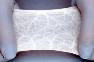
3D Cellular Matrix Scaffold: Providing an Ideal Environment for Healing and Cartilage Repair
A crucial part of our treatment strategy involves a state-of-the-art 3D cellular matrix scaffold rich in platelet lysate. This scaffold provides an ideal environment for the chondrocytes and Hypo-MSCs to grow and function. It’s a concept inspired by nature itself – just as cells in our body reside in a structured environment, this 3D scaffold serves as a ‘home’ for the cells, giving them the support and nutrients they need to thrive.
But what makes our 3D scaffold so unique? It’s made from platelet lysate, a substance derived from platelets rich in growth factors and proteins beneficial for cell growth and tissue repair. This environment simulates the natural conditions that cells need to flourish, enhancing our ChondRX™ product’s efficacy. In addition to providing physical support, the scaffold also ensures that the cells remain at the injury site, maximizing their regenerative potential.
Existing research echoes the potential benefits of using a scaffold in cartilage repair. For example, a study conducted by Chang et al. explored the use of a silk fibroin scaffold for cartilage tissue engineering and found promising results [10].
Our approach extends this concept further by employing the scaffold as a delivery method for chondrocytes and Hypo-MSCs via injection rather than surgical implantation. This non-surgical approach offers the benefits of a less invasive procedure with potentially quicker recovery times.
A Synergistic Approach to Cartilage Repair: ChondRX™, Hypo-MSCs, and 3D Scaffold
The power of ChondRX™ lies in the unique synergy of its components: the chondrocytes, Hypo-MSCs, and the 3D cellular matrix scaffold. Each component has a unique role, but they all work together to amplify the healing and regenerative potential.
Chondrocytes form and maintain cartilage, generating collagen and other components crucial for the structural integrity of our joints. Hypo-MSCs, with their heightened potency, promote tissue regeneration, combat inflammation, and enhance angiogenesis. In addition, the 3D cellular matrix scaffold offers a conducive environment for these cells to thrive, ensuring they stay at the site of injury for optimal healing.
In essence, ChondRX™ represents a monumental stride in regenerative medicine. It merges the advantages of chondrocytes, the amplified power of Hypo-MSCs, and the supportive environment of the 3D cellular matrix scaffold to offer a revolutionary solution for joint and disc injuries.
At CPI, we are proud to be the pioneers of this advanced treatment strategy. We are excited about the potential of ChondRX™ and its positive implications for our patients. We remain committed to improving lives through advanced science and technology as we continue our innovation journey.
If you or a loved one is interested in using stem cells for cartilage repair we offer a free doctor’s phone consultation Please click here to book your free call.
**Citations**:
[1] Sophia Fox, A.J., Bedi, A., & Rodeo, S.A. (2009). The Basic Science of Articular Cartilage: Structure, Composition, and Function. Sports Health, 1(6), 461–468.
https://www.ncbi.nlm.nih.gov/pmc/articles/PMC3445147/
[2] Eyre, D. (2004). Collagens and cartilage matrix homeostasis. Clinical Orthopaedics and Related Research (1976-2007), 427(Suppl), S118-S122.
https://pubmed.ncbi.nlm.nih.gov/15480053/
[3] Brittberg, M., et al. (1994). Treatment of Deep Cartilage Defects in the Knee with Autologous Chondrocyte Transplantation. New England Journal of Medicine, 331(14), 889-895.
https://pubmed.ncbi.nlm.nih.gov/8078550/
[4] Vasiliadis, H. S., & Wasiak, J. (2010). Autologous Chondrocyte Implantation for Full Thickness Articular Cartilage Defects of the Knee. Cochrane Database of Systematic Reviews, Issue 10. Art. No.: CD003323.
https://www.ncbi.nlm.nih.gov/pmc/articles/PMC7144735/
[5] Tsai, C.C., et al. (2011). Hypoxia inhibits senescence and maintains mesenchymal stem cell properties through down-regulation of E2A-p21 by H
https://europepmc.org/article/med/20952688
[6] Hu, X., et al. (2016). Hypoxia preconditioning enhances the viability of ADSCs to increase the survival rate of ischemic skin flaps in rats. Aesthetic Plastic Surgery, 40(6), 892-901.
https://pubmed.ncbi.nlm.nih.gov/23232730/
[7] Beegle, J., et al. (2015). Hypoxic Preconditioning of Mesenchymal Stromal Cells Induces Metabolic Changes, Enhances Survival, and Promotes Cell Retention In Vivo. Stem Cells, 33(6), 1818-1828.
https://pubmed.ncbi.nlm.nih.gov/25702874/
[8] Liu, H., et al. (2012). Hypoxia preconditioned human adipose-derived mesenchymal stem cells enhance angiogenic potential via secretion of increased VEGF and bFGF. Cell Biology International, 36(6), 493-502.
https://pubmed.ncbi.nlm.nih.gov/23505143/
[9] JF Silva, et al. (2018) Comparing atmospheric and hypoxic cultured mesenchymal stem cell transcriptome: implication for stem cell therapies targeting intervertebral discs
https://pubmed.ncbi.nlm.nih.gov/30097061/
[10] Chang, S., et al. (2013). The in vivo performance of plasmin-sensitive RGD-alginate scaffolds containing basic fibroblast growth factor. Biomaterials, 34(4), 1011-1018.